

Palpation of the lateral pterygoid has been the subject of several discussions, and these generate many controversies, precisely due to the topography of the muscle. Therefore, knowing whether it is possible to perform digital palpation would be of great clinical value in the investigation of various problems that affect the temporomandibular joint.
To answer this question, Wolfgang et al. (2015), carried out excellent research, which consisted of performing an MRI on a 30-year-old man, to observe the location of the lateral pterygoid (Figure 1), after which the examiner performed digital palpation with the little finger or with the index finger, and during palpation a new magnetic resonance examination was performed (Figure 2).

Figure 1 - Palpation without opening the mouth.
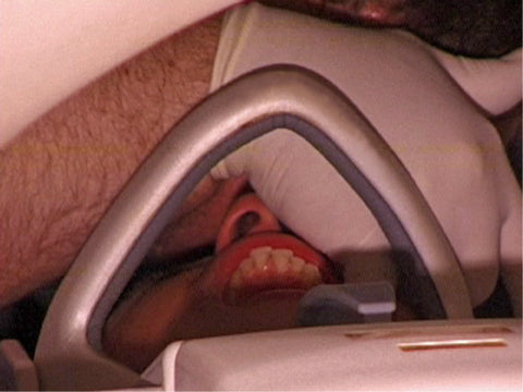
Figure 2 - Palpation with mouth opening, during magnetic resonance imaging.
Figure 3 shows a 6mm deformation that occurred in the muscle during palpation with the index finger during magnetic resonance imaging. This way, it is possible to report that it is possible to palpate the lateral pterygoid. 
Figure 3 - Red outline: lateral pterygoid muscle; yellow outline: examiner's finger. Magnetic resonance image demonstrating palpation of the lateral pterygoid muscle.
The technique used to palpate is already well described and used, to show whether it is effective, the authors decided to touch the lateral pterygoid in five cadavers, observing whether the touch occurred.
During this test with cadavers, it was seen that all digital touches reached the target muscle, after exposing the infratemporal fossa, as can be seen in figures 4 and 5.

Figure 4 - Palpation of the target muscle in cadavers.

Figure 5 - Digital reaching the lateral pterygoid. (yellow dot: medial pterygoid; red dot: lateral pterygoid).
To perform this palpation successfully, it is worth remembering that the patient must perform the ipsilateral lateroprotrusion movement at the time of palpation, as if the muscle is relaxed, palpation may be impaired.

Figure 6 - Patient performing ipsilateral lateroprotrusion movement before beginning palpation.
The lateral pterygoid muscle is key to the treatment of temporomandibular disorders (TMD), since dysfunction of this muscle is one of the main causes of oral pain. Now, with the possibility of performing palpation clearly, clinical analyzes are more precise.
Click on the link below to access the original article: